Radiologie des kindlichen Thorax
Didaktisches Konzept:
- Das Schwergewicht von PediRad liegt auf dem Bild. Theoretisches Wissen holen Sie sich besser aus Büchern und in Vorlesungen.
- PediRad stellt bilddiagnostisches Übungsmaterial aus dem Gebiet der Kinderradiologie für Studenten und Assistenzärzte verschiedener Fachgebiete zur Verfügung.
- Die Übungseinheit „Thoraxdiagnostik“ stellt umfangreiches radiologisches Bildmaterial zu normalen und pathologischen Situationen zur Verfügung; im Zentrum steht dabei das klassische konventionelle Röntgenbild.
- Die korrekte Interpretation des „Röntgenthorax“ in zwei Ebenen stellt eine der wichtigsten Grundlagen für eine erfolgreiche und verantwortungsvolle klinische Tätigkeit dar.
- Herkömmliche Lehrmedien können aus Platz- und Kostengründen jeweils nur die ganz normalen gesunden und sehr typischen pathologischen Bilder wiedergeben. Das ist für Lernende bei weitem nicht ausreichend. Die Spielarten des Normalen und Pathologischen sind häufiger als das Emblematische der „Textbooks“.
- Mit dem „Picturebook“ wollen wir durch Vielfalt und Spielarten Vergleichsmöglichkeiten eröffnen, die einen klinikbezogenen Wissenserwerb ermöglichen und den Vorsprung des Erfahrenen vielleicht etwas schrumpfen lassen.
Lernziele dieses Lernprogramms:
- Einsicht in die Vielfalt und die Spielarten des Normalen und Pathologischen.
- Nachvollziehen der Entwicklungsschritte des kindlichen Thorax anhand der radiologischen Manifestationen.
- Erkennen eines normalen Befundes in Abhängigkeit des Alters der kleinen und größeren Patienten.
- Erkennen eines normalen Befundes in Abhängigkeit von speziellen Situationen (Atemlage, Körperposition, Intubation, etc.).
- Nachvollziehen der Charakteristika pathologischer Befunde verschiedener Ausprägung.
- Erarbeitung des Stoffes nach unterschiedlichen Systematiken (anatomische Regionen, pathologische Gruppen, Altergruppen)
- Quervergleiche innerhalb des Normalen, innerhalb des Pathologischen und zwischen den beiden Gruppen.
- Sammeln und Nacharbeiten von Bildern für individuelle Zwecke mittels des Bildarchivs.
- Üben von klinischen Situationen aus dem täglichen Leben anhand der Fallbeispiele.
Beteiligte Personen:
- Jan Nagy, med. pract., Erarbeitung des Stoffes und Aufbereitung der Fälle.
- Dr. Rainer Wolf, Leitender Arzt, Pädiatrische Radiologie, fachliche Betreuung.
- Dr. Ulrich Woermann, didaktische Betreuung, Projektleitung.
- M. Rolli, med. pract., Entwicklungsdatenbank.
Chest radiography in children
Didactical concept:
- The emphasis of PediRad is on the plain radiograph. Theoretical knowledge should be achived from books and in lectures.
- PediRad makes radiograph-diagnostic exercise material available from the area of child radiology for students and assistant physician of different fields of activity.
- The exercise unit "chest diagnostics" offers extensive radiological pictorial material to normal and pathological situations; in the centre thereby the classical plain or conventional radiograph is located.
- The correct interpretation and evaluation of the "chest radiograph" in two levels represents one of the most important basis for a successful and responsible clinical activity.
- Conventional training media can show, because of place and cost reasons, only the completely normal and very typical pathological radiographs. That is certainly not sufficient for learners. Variations of the "normal" and "pathological" are far more frequent than examples shown in teaching books.
- With "the picturebook" we want to show comparison possibilities, which enable you to achive good clinical knowledge and experience.
Learning objectives of this teaching program:
- Insight into the variety of "normal" and the "pathological".
- Ability to reconstruct the developmental steps of chest in children on the basis of radiological manifestations.
- Recognise "normal findings" depending on the age of the smaller and bigger patients.
- Recognise "normal findings" as a function of special situations (inspiration situation, body position, intubation, etc.).
- Understand the characteristics of pathological findings of different developments.
- To compile the material by different systems (anatomical regions, pathological groups, age groups)
- Comparisons within the "normal findings", within the "pathological findings" and between the two groups.
- Collecting and re-working pictures for individual purposes by means of picture archives.
- Practice clinical situations from the daily life on the basis of the case examples.
People involved in this project:
- Jan Nagy MD, development, editing of the cases and translation into English.
- Caroline Phillipson MBBS, translation into English
- Rainer Wolf MD, consultant, head of Pediatric Radiology, technical support
- Ulrich Woermann MD, didactical support, direction of the project.

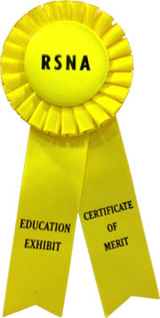
Die englische Version von PediRad Thorax erhielt das Certificate of Merit an der 97. wissenschaftlichen Jahresversammlung der Radiological Society of North America RSNA 2011 in Chicago. PediRad Chest was awarded the Certificate of Merit at the 97th Scientific Assembly and Annual Meeting of the Radiological Society of North America 2011 in Chicago.